Where are the digestive cells of hydra located? Hydras
Tamara Yakovlevna’s book, published in 2003, was written by a person who studied this issue in practice for many years. For our topic, it is especially interesting because it provides a detailed classification of those cells that are found in human blood. I will not waste time on a detailed retelling of the content and provide this detailed classification here. Anyone can read it for themselves. I will note only the main points.
Firstly, when describing many types of blood cells, the phrase “functional role is not well understood” is used.
Secondly, it turns out that there are many types of cells that are similar in appearance, but differ in internal structure. Including those that have a different shape and structure of the cell nucleus, as well as the presence or absence of various internal structural elements.
Thirdly, in the process of her research, Tamara Yakovlevna came to the conclusion that some of the microorganisms, for example the same Trichomonas, can take different forms, including mimicking, becoming similar to lymphocytes! At the same time, she came to this conclusion largely because some of these false “lymphocytes” behaved like Trichomonas, destroying and devouring blood cells, primarily red blood cells, which is also observed in Trichomonas in the blood.
Hypothesis for what "hydra" is needed

In my opinion, it is quite obvious that the lymphatic system is not a self-sufficient organism that could exist outside the host’s body. It has neither an internal skeleton, nor an external strong protective shell, nor many different organs, without which no independent organism can exist. If we consider the lymphatic system as a certain independent entity, then this structure has at least some meaning only when it is built into the human body and uses the organs of the body for its needs. Therefore, there is no point in “teleporting” it anywhere or moving it in any other way without the rest of the body. Also confusing is the very low efficiency of the system we see on Earth today. Out of almost 7 billion living people, the state in which, according to Konstantin, the maturation and “teleportation” of a mature individual of “hydra” occurs, reaches at most several hundred people. This is too little, especially considering that Constantine considers the “hydra” to be an intelligent entity. If she were truly intelligent, and she really needed mature new individuals, then the entire system would be built in such a way as to provide as many mature individuals as possible. When I asked this question to Konstantin, he answered the following: “Regarding efficiency. I don’t see anything surprising in such efficiency. If you take the percentage of all grains of one type of tree that fall in the forest, then exactly the same percentage will reach full maturity.” Alas, the argument is not very convincing, since either the “hydra” does not exceed the plant’s intellectual abilities, then we can reluctantly accept such a low efficiency, or the “hydra” itself or its creator and owner has a mind, and a very advanced one, if he managed to capture and enslave a planet with a fairly highly developed civilization that did not live on it. But then such a low efficiency clearly indicates that the meaning of this idea is completely different.
I thought about this topic for quite a long time, and ultimately came to the following conclusions. If we continue the analogy with technical systems, then in the same racing cars, when boosting the engine in order to increase its peak power, among other things, they strengthen the exhaust system, which will remove additional combustion products when operating at increased power. The exhaust channels are made larger to reduce resistance to escaping gases, and additional exhaust pipes are added. At the same time, such a modified engine actually increases power. But at the same time, he also has a very serious drawback! When we transfer the engine to work with increased power, for which its design was not originally designed, this very sharply reduces its service life. Usually by several times. In other words, such a modification shortens lifespan!
We see exactly the same thing in the case of the human body. Our body was equipped with an additional excretory system, since something was changed in the general principle of its functioning. At the same time, available facts indicate that this system operates constantly and does not turn on only during illness or damage to the body. That is why, when a serious illness occurs or the body receives a serious injury, its capabilities are not enough to remove all the resulting toxins from the body, since it is already loaded.
So the “hydra”, by its purpose, is a suppression system, which, firstly, blocks part of the mental abilities, suppressing some functions of the nervous system and brain, including having a chemical effect on it. Secondly, it shortens lifespan due to disruption of internal metabolic processes, which I will discuss in more detail below. And, finally, thirdly, it sharply reduces the overall energy potential of a person, since the body is forced to constantly spend energy on restoring the cells destroyed by the “hydra”.
Here it is necessary to mention one more fact, which has been confirmed experimentally. The fact is that the total number of cell divisions is not infinite. There is a so-called limit or Hayflick limit, which is associated with the peculiarity of copying a DNA molecule during cell division. It is believed that the copying process occurs in such a way that the outermost portions of the ends of the DNA, which is usually shaped like the letter X, can be copied. That is, it is somewhat similar to printing on some printers, where they inevitably leave margins of white paper at the edges where the feed rollers grab the sheet as they pull it through the printing mechanism.

I also learned that today some researchers already have doubts about what causes the shortening of telomeres (the ends of the DNA molecule), but at the same time everyone agrees that it is the achievement of the ends of the telomeres of a certain minimum value that leads to the cessation of cell division and its subsequent apoptosis. Apoptosis is a natural process of cell death and resorption in the body, which is distinguished from necrosis - cell death due to an external negative process.
A separate question to which I could not find a definite answer is the lifespan of cells of various human tissues. A variety of terms are given, from 120 days to 15 years. Moreover, the first period of 120 days I heard in a program on Vesti FM radio, dedicated to the topic of health, where some female doctor spoke (unfortunately, I did not hear who exactly). But this period is clearly not true, since with a limit of 52 divisions, the total lifespan of the organism will be only 6,240 days or a little more than 17 years, and from the moment of conception of the fetus. And if we take into account that in the initial period of development of the body, cell division occurs much more often, approximately once a day, then if Hayflick’s theory is correct, the embryo will have to die on the 52nd day after conception. And since this does not happen, we can once again repeat the same phrase “the mechanism of cell functioning has not been sufficiently studied.” Obviously, there must be some other mechanism that generates new cells with complete DNA. Most likely, the spinal cord and thymus (thymus gland) are somehow involved in this process, but this is a topic for another article. In addition, it has already been proven that the Hayflick limit does not manifest itself in a number of cells, including cancer cells, which can divide almost an unlimited number of times.
However, since the presence of the Hayflick limit for most somatic cells has been established and confirmed experimentally, we will assume that after the cell matures and differentiates, when it takes its permanent position in the body, it actually turns on a certain mechanism that limits the number of divisions . This in turn means that if the process that generates new cells does so more slowly than the mature cells in the body age and die, then the lifespan of such an organism will be limited.
What do we know from the mythology of different nations, including from the same Bible. Once upon a time, a person was immortal or lived for a very long time, according to the Bible about 1000 years. The chart below shows the ages of the key characters in the Old Testament.
Why shorten a person's life? This does not allow us to gain the intellectual potential that we should have. Back in school we were told that most people use no more than 10% of their brain capacity. But we cannot use its capabilities 100% if today we actually live less than 10% of the period that we should live based on the potential of our body. That is, we don’t use all the opportunities not because we don’t want to, but because we simply don’t have time to use them. We do not have time to form in our brain a neural network of such complexity and quality that would allow us to fully understand the processes occurring around us in order to fully and effectively manage them. We only look adults outwardly, but intellectually, compared to what we should be, we remain underdeveloped children. This is very convenient for the invaders of our planet, since children with undeveloped intelligence are much easier to deceive and keep under control.
So, at the moment, for myself personally, I have come to the following conclusions.
Intense cell destruction causes the remaining cells to divide more frequently to make up for the losses. At the same time, the existing process of generating new cells, which have the full length of telomeres in DNA molecules, does not have time to form the required number of new cells to renew all tissues of the body. That is why the tissues of our body begin to age and become decrepit gradually, and not all at once. In a young organism, all tissue cells are still young. They begin to be destroyed by the "hydra", forcing the remaining cells to divide. Over time, an increasing number of cells become old, since the mechanism for synthesizing new young cells does not have time to produce the required number of cells to replace all the dying ones. That is, old, decrepit tissue differs from young tissue precisely in that the percentage of cells in it that have already reached the limit of division and begun to degrade is higher than new cells.
Accordingly, if the body is subjected to some additional destructive influence, for example while working in a hazardous industry, this will lead to even more rapid cell death. Therefore, the body tissues of such a person will age and become decrepit much faster than those of someone who is not exposed to such additional harmful effects. This also includes many other destructive factors, ranging from poor ecology to alcoholism. Each of you can easily find proof of this fact around you.
To be continued...
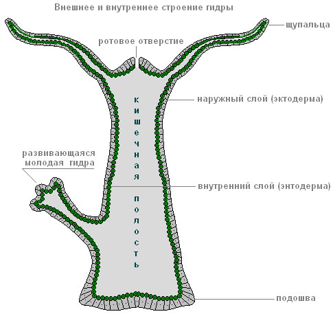
Figure: Structure of freshwater hydra. Radial symmetry of Hydra
Habitat, structural features and vital functions of the freshwater hydra polyp
In lakes, rivers or ponds with clean, transparent water, a small translucent animal is found on the stems of aquatic plants - polyp hydra(“polyp” means “multi-legged”). This is an attached or sedentary coelenterate animal with numerous tentacles. The body of an ordinary hydra has an almost regular cylindrical shape. At one end is mouth, surrounded by a corolla of 5-12 thin long tentacles, the other end is elongated in the form of a stalk with sole at the end. Using the sole, the hydra is attached to various underwater objects. The body of the hydra, together with the stalk, is usually up to 7 mm long, but the tentacles can extend several centimeters.
Radial symmetry of Hydra
If you draw an imaginary axis along the body of the hydra, then its tentacles will diverge from this axis in all directions, like rays from a light source. Hanging down from some aquatic plant, the hydra constantly sways and slowly moves its tentacles, lying in wait for prey. Since the prey can appear from any direction, the tentacles arranged in a radial manner are best suited to this method of hunting.
Radiation symmetry is characteristic, as a rule, of animals leading an attached lifestyle.
Hydra intestinal cavity
The body of the hydra has the form of a sac, the walls of which consist of two layers of cells - the outer (ectoderm) and the inner (endoderm). Inside the body of the hydra there is intestinal cavity(hence the name of the type - coelenterates).
The outer layer of hydra cells is the ectoderm.

Figure: structure of the outer layer of cells - hydra ectoderm
The outer layer of hydra cells is called - ectoderm. Under a microscope, several types of cells are visible in the outer layer of the hydra - the ectoderm. Most of all here are skin-muscular. By touching their sides, these cells create the cover of the hydra. At the base of each such cell there is a contractile muscle fiber, which plays an important role in the movement of the animal. When everyone's fiber skin-muscular cells contract, the hydra's body contracts. If the fibers contract on only one side of the body, then the hydra bends in that direction. Thanks to the work of muscle fibers, the hydra can slowly move from place to place, alternately “stepping” with its sole and tentacles. This movement can be compared to a slow somersault over your head.
The outer layer contains and nerve cells. They have a star-shaped shape, as they are equipped with long processes.
The processes of neighboring nerve cells come into contact with each other and form nerve plexus, covering the entire body of the hydra. Some of the processes approach the skin-muscle cells.
Hydra irritability and reflexes
Hydra is able to sense touch, temperature changes, the appearance of various dissolved substances in water and other irritations. This causes her nerve cells to become excited. If you touch the hydra with a thin needle, then the excitement from irritation of one of the nerve cells is transmitted along the processes to other nerve cells, and from them to the skin-muscle cells. This causes muscle fibers to contract, and the hydra shrinks into a ball.

Picture: Hydra's irritability
In this example, we get acquainted with a complex phenomenon in the animal body - reflex. The reflex consists of three successive stages: perception of irritation, transfer of excitation from this irritation along the nerve cells and response body by any action. Due to the simplicity of the hydra's organization, its reflexes are very uniform. In the future we will become familiar with much more complex reflexes in more highly organized animals.
Hydra stinging cells

Pattern: Stringing or nettle cells of Hydra
The entire body of the hydra and especially its tentacles are seated with a large number stinging, or nettles cells. Each of these cells has a complex structure. In addition to the cytoplasm and nucleus, it contains a bubble-like stinging capsule, inside which a thin tube is folded - stinging thread. Sticking out of the cage sensitive hair. As soon as a crustacean, small fish or other small animal touches a sensitive hair, the stinging thread quickly straightens, its end is thrown out and pierces the victim. Through a channel passing inside the thread, poison enters the body of the prey from the stinging capsule, causing the death of small animals. As a rule, many stinging cells are fired at once. Then the hydra uses its tentacles to pull the prey to its mouth and swallows it. The stinging cells also serve the hydra for protection. Fish and aquatic insects do not eat hydras, which burn their enemies. The poison from the capsules is reminiscent of nettle poison in its effect on the body of large animals.
The inner layer of cells is the hydra endoderm

Figure: structure of the inner layer of cells - hydra endoderm
Inner layer of cells - endoderm A. The cells of the inner layer - the endoderm - have contractile muscle fibers, but the main role of these cells is to digest food. They secrete digestive juice into the intestinal cavity, under the influence of which the hydra’s prey softens and breaks down into small particles. Some of the cells of the inner layer are equipped with several long flagella (as in flagellated protozoa). The flagella are in constant motion and sweep particles towards the cells. The cells of the inner layer are capable of releasing pseudopods (like those of an amoeba) and capturing food with them. Further digestion occurs inside the cell, in vacuoles (like in protozoa). Undigested food remains are thrown out through the mouth.
The hydra has no special respiratory organs; oxygen dissolved in water penetrates the hydra through the entire surface of its body.
Hydra regeneration
The outer layer of the hydra's body also contains very small round cells with large nuclei. These cells are called intermediate. They play a very important role in the life of the hydra. With any damage to the body, intermediate cells located near the wounds begin to grow rapidly. From them, skin-muscle, nerve and other cells are formed, and the wounded area quickly heals.
If you cut a hydra crosswise, tentacles grow on one of its halves and a mouth appears, and a stalk appears on the other. You get two hydras.
The process of restoring lost or damaged body parts is called regeneration. Hydra has a highly developed ability to regenerate.
Regeneration, to one degree or another, is also characteristic of other animals and humans. Thus, in earthworms it is possible to regenerate a whole organism from their parts; in amphibians (frogs, newts) entire limbs, various parts of the eye, tail and internal organs can be restored. When a person is cut, the skin is restored.
Hydra reproduction
Asexual reproduction of hydra by budding

Figure: Hydra asexual reproduction by budding
Hydra reproduces asexually and sexually. In summer, a small tubercle appears on the hydra’s body - a protrusion of the wall of its body. This tubercle grows and stretches out. Tentacles appear at its end, and a mouth breaks out between them. This is how the young hydra develops, which at first remains connected to the mother with the help of a stalk. Outwardly, all this resembles the development of a plant shoot from a bud (hence the name of this phenomenon - budding). When the little hydra grows up, it separates from the mother’s body and begins to live independently.
Hydra sexual reproduction
By autumn, with the onset of unfavorable conditions, hydras die, but before that, sex cells develop in their body. There are two types of germ cells: ovoid, or female, and spermatozoa, or male reproductive cells. Sperm are similar to flagellated protozoa. They leave the hydra's body and swim using a long flagellum.

Figure: Hydra sexual reproduction
The hydra egg cell is similar to an amoeba and has pseudopods. The sperm swims up to the hydra with the egg cell and penetrates inside it, and the nuclei of both sex cells merge. Happening fertilization. After this, the pseudopods are retracted, the cell is rounded, and a thick shell is formed on its surface - a egg. At the end of autumn, the hydra dies, but the egg remains alive and falls to the bottom. In the spring, the fertilized egg begins to divide, the resulting cells are arranged in two layers. From them a small hydra develops, which, with the onset of warm weather, comes out through a break in the egg shell.
Thus, the multicellular animal hydra at the beginning of its life consists of one cell - an egg.
The hydra's body looks like an oblong sac, the walls of which consist of two layers of cells - ectoderm And endoderm.
Between them lies a thin gelatinous non-cellular layer - mesoglea, serving as a support.
The ectoderm forms the covering of the animal’s body and consists of several types of cells: epithelial-muscular, intermediate And stinging.
The most numerous of them are epithelial-muscular.
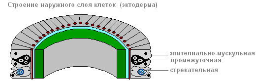
Ectoderm

epithelial muscle cell
Due to muscle fibers, lying at the base of each cell, the body of the hydra can contract, lengthen and bend.
Between the epithelial-muscle cells there are groups of small, round cells with large nuclei and a small amount of cytoplasm, called intermediate.

When the hydra's body is damaged, they begin to grow and divide rapidly. They can transform into other types of cells in the hydra body, except for epithelial-muscular ones.
The ectoderm contains stinging cells, serving for attack and defense. They are mainly located on the tentacles of the hydra. Each stinging cell contains an oval capsule in which the stinging filament is coiled.

Structure of a stinging cell with a coiled stinging thread
If prey or an enemy touches a sensitive hair located outside the stinging cell, in response to irritation the stinging thread is ejected and pierces the body of the victim.
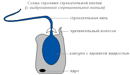
Structure of a stinging cell with discarded stinging thread
Through the thread channel, a substance that can paralyze the victim enters the victim’s body.
There are several types of stinging cells. The threads of some pierce the skin of animals and introduce poison into their bodies. The threads of others are wrapped around the prey. The threads of the third are very sticky and stick to the victim. Usually the hydra “shoots” several stinging cells. After the shot, the stinging cell dies. New stinging cells are formed from intermediate.
The structure of the inner layer of cells
Endoderm lines the entire intestinal cavity from the inside. It includes digestive-muscular And glandular cells.

Endoderm
Digestive system
There are more digestive muscle cells than others. Muscular fibers they are capable of reduction. When they shorten, the hydra's body becomes thinner. Complex movements (movement by “tumbling”) occur due to contractions of muscle fibers of ectoderm and endoderm cells.
Each of the digestive-muscle cells of the endoderm has 1-3 flagella. Hesitating flagella create a current of water, which drives food particles towards the cells. Digestive-muscle cells of the endoderm are capable of forming pseudopods, capture and digest small food particles in the digestive vacuoles.
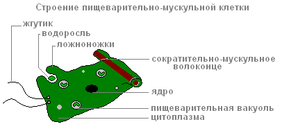
The structure of the digestive muscle cell
Glandular cells in the endoderm secrete digestive juice into the intestinal cavity, which liquefies and partially digests food.
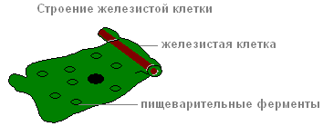
The structure of the glandular cell
Prey is captured by the tentacles using stinging cells, the venom of which quickly paralyzes small victims. By coordinated movements of the tentacles, the prey is brought to the mouth, and then, with the help of body contractions, the hydra is “put on” the victim. Digestion begins in the intestinal cavity ( cavity digestion), ends inside the digestive vacuoles of epithelial-muscular endoderm cells ( intracellular digestion). Nutrients are distributed throughout the hydra's body.
When the digestive cavity contains remains of the prey that cannot be digested, and waste products of cellular metabolism, it contracts and empties.
Breath
Hydra breathes oxygen dissolved in water. She has no respiratory organs, and she absorbs oxygen over the entire surface of her body.
Circulatory system
Absent.
Selection
The release of carbon dioxide and other unnecessary substances formed during life processes is carried out from the cells of the outer layer directly into the water, and from the cells of the inner layer into the intestinal cavity, then out.
Nervous system
Below the skin-muscle cells are star-shaped cells. These are nerve cells (1). They connect with each other and form a nerve network (2).

Nervous system and irritability of the hydra
If you touch the hydra (2), then excitation (electrical impulses) occurs in the nerve cells, which instantly spreads throughout the entire nervous network (3) and causes contraction of the skin-muscle cells and the entire body of the hydra shortens (4). The response of the hydra body to such irritation is unconditioned reflex.
Sex cells
With the approach of cold weather in the fall, germ cells are formed from intermediate cells in the ectoderm of the hydra.
There are two types of germ cells: eggs, or female germ cells, and sperm, or male germ cells.
The eggs are located closer to the base of the hydra, sperm develop in tubercles located closer to the mouth.
egg cell Hydra is similar to an amoeba. It is equipped with pseudopods and grows rapidly, absorbing neighboring intermediate cells.
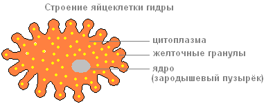
The structure of the hydra egg cell

The structure of the hydra sperm
Sperm in appearance they resemble flagellated protozoa. They leave the hydra's body and swim using a long flagellum.
Fertilization. Reproduction
The sperm swims up to the hydra with the egg cell and penetrates inside it, and the nuclei of both sex cells merge. After this, the pseudopods are retracted, the cell is rounded, a thick shell is released on its surface - an egg is formed. When the hydra dies and is destroyed, the egg remains alive and falls to the bottom. With the onset of warm weather, the living cell located inside the protective shell begins to divide, the resulting cells are arranged in two layers. From them a small hydra develops, which comes out through a break in the egg shell. Thus, the multicellular animal hydra at the beginning of its life consists of only one cell - an egg. This suggests that the ancestors of Hydra were single-celled animals.

Asexual reproduction of hydra
Under favorable conditions, hydra reproduces asexually. A bud forms on the animal’s body (usually in the lower third of the body), it grows, then tentacles form and a mouth breaks through. The young hydra buds from the mother's body (in this case, the mother and daughter polyps are attached with tentacles to the substrate and pull in different directions) and leads an independent lifestyle. In autumn, hydra begins to reproduce sexually. On the body, in the ectoderm, gonads are formed - sex glands, and in them, germ cells develop from intermediate cells. When hydra gonads form, a medusoid nodule is formed. This suggests that the hydra gonads are highly simplified sporifers, the last stage in the series of transformation of the lost medusoid generation into an organ. Most species of hydra are dioecious; hermaphroditism is less common. Hydra eggs grow rapidly by phagocytosing surrounding cells. Mature eggs reach a diameter of 0.5-1 mm. Fertilization occurs in the body of the hydra: through a special hole in the gonad, the sperm penetrates the egg and merges with it. The zygote undergoes complete uniform fragmentation, as a result of which a coeloblastula is formed. Then, as a result of mixed delamination (a combination of immigration and delamination), gastrulation occurs. A dense protective shell (embryotheca) with spine-like outgrowths is formed around the embryo. At the gastrula stage, the embryos enter suspended animation. Adult hydras die, and the embryos sink to the bottom and overwinter. In the spring, development continues, in the parenchyma of the endoderm, an intestinal cavity is formed by divergence of cells, then the rudiments of tentacles are formed, and a young hydra emerges from under the shell. Thus, unlike most marine hydroids, hydra does not have free-swimming larvae and its development is direct.
Regeneration
Hydra has a very high ability to regenerate. When cut crosswise into several parts, each part restores the “head” and “leg”, maintaining the original polarity - the mouth and tentacles develop on the side that was closer to the oral end of the body, and the stalk and sole develop on the aboral side of the fragment. The whole organism can be restored from individual small pieces of the body (less than 1/100 of the volume), from pieces of tentacles, and also from a suspension of cells. Moreover, the regeneration process itself is not accompanied by increased cell division and is a typical example of morphallaxis.
Movement
In a calm state, the tentacles extend several centimeters. The animal slowly moves them from side to side, lying in wait for prey. If necessary, the hydra can move slowly.
"Walking" mode of transportation

"Walking" method of movement of the hydra
Having curved its body (1) and attached its tentacles to the surface of an object (substrate), the hydra pulls the sole (2) to the front end of the body. Then the walking movement of the hydra is repeated (3,4).
"Tumbling" mode of movement

"Tumbling" method of movement of the hydra
In another case, it seems to tumble over its head, alternately attaching itself to objects with its tentacles and its sole (1-5).
- lymph is a foreign full-fledged organism in the human body
- circulatory and lymphatic systems
- what in the body is attacked by viruses
- immunity, incubation
- encephalitis is a harmless infection, but...
Dmitry Mylnikov
Hydra. Part 1a

And here is another image, there are a hundred years between the engravings.

If we collect the basic information that we managed to collect on Hydra, we get:
1. The Hydra's body occupies the entire intercellular space, swaddling the body from the outside, occupying energy channels, the excretory system of the skin (pores) and all places where there is no obvious blood flow, where there is no strong immunity.
2. Hydra's various internal organs are located throughout the body.
3. The bulk of the hydra’s body and its center are located in the intestinal cavity. There are also larvae or heads there. Official science believes that 90 percent of immunity is spent in the digestion process.
The main conclusion: in the human body, “Hydra” is represented by the human lymphatic system, and lymphocytes are the cells of “Hydra”. At the same time, the lymphatic vessels are the “circulatory” system of the Hydra body. It does not intersect with the circulatory system of our body and there is no immunity in it other than the immunity of Hydra itself.


The lymphatic system, with its thin capillaries, permeates the entire structure of the body. Its main functions are to conduct lymph from tissues into the venous bed; absorption from the intercellular space of colloidal solutions of protein substances that are not absorbed into the blood capillaries; absorption of water and crystalloids dissolved in it; the formation of lymphocytes involved in immunological reactions, and the neutralization of foreign particles, microbes, and bacteria entering the body.”
Here I would like to draw your attention to the fact that such an important system of the body is not studied in medical universities! I hope that after reading this material you will understand why. But let’s return to the above description and try to understand what’s really wrong there.
Firstly, it is argued that the task of lymphatic vessels is to absorb colloidal solutions of protein substances, which are supposedly not absorbed into the capillaries of blood vessels. Moreover, the diameter of both is actually the same, and there is water in both. The reason why these colloidal solutions should not be absorbed into the capillaries of the circulatory system, but are absorbed into the capillaries of the lymphatic system, is not explained. They don’t get absorbed and that’s it. But the most important thing is that the substances collected by the lymphatic system are not removed outward, but back into the blood, since the lymphatic vessels ultimately enter the venous bed! This means that further removal of all these toxins and breakdown products is still carried out by our kidneys and liver!
Secondly, the lymphatic system has one important drawback. Unlike the circulatory system, which has its own heart-shaped pump that creates a constant flow of blood, the lymphatic system does not have its own pump! Lymph moves through the lymphatic vessels due to the valves located in them and their constant compression and expansion during muscle contraction.

But if the lymphatic system is part of the immune system, which must remove toxins and harmful substances from the body, then this design has a very serious drawback, because when the body gets sick, its mobility is minimal, since it begins to spend most of its energy on fighting the disease . It turns out that it is at this moment that the lymphatic system actually does not really function! How so? But what about the removal of harmful substances from the body, the same colloidal solutions? How does the body remove them from the body during illness, when its mobility is minimal? And why don’t we die from intoxication?
The portal “Sedition” recently published a very interesting . When you read this article for the first time, it seems that you are learning something new and important about how our body works and works. But this is only until you begin to analyze its content.
A comment rodline about the study of lymph and stitching of lymph vessels:
I asked around. They study - the material is presented in several disciplines, i.e., from different sides. Interaction mechanisms, device, etc..
During operations, large vessels are still sutured. Small ones - no.
Well, it probably also depends on the conscientiousness of the surgeon.
A comment dobrosvet108 about DNA:
These geneticists are complete fools, so what they do not understand is equated to garbage. Only beings with a low spiritual level, although with a developed mind, can do this. The state of modern official science is no better than the state of society as a whole. So you can’t rely on this in any way. DNA is full of multidimensional information, there is nothing superfluous.
Animals. Hydras live in stagnant or slowly flowing water - in ponds, lakes and river backwaters rich in aquatic vegetation. The freshwater hydra is a very small animal, measuring 1 - 3 cm in size, so the hydra is difficult to detect. It is best to observe the life of hydras using a magnifying glass or microscope. The easiest way to find a hydra is to scoop water from a pond into a jar along with duckweed floating on the surface. If such water is allowed to settle, then on the wall of the vessel facing the light you will notice small, thin tubes of white, brown or green color. These are freshwater hydras. There are several types of freshwater hydra.
Nutrition
Freshwater hydra feeds on small aquatic animals. Place tiny animals from the pond - crustaceans, daphnia, or water fleas - into a jar or aquarium in which freshwater hydras live, and you will see how important tentacles are in the life of a hydra. If any daphnia, moving quickly in the water, slightly touches the tentacles of the hydra, it stops as if paralyzed and completely loses the ability to move. The hydra's tentacles envelop the daphnia and pull it towards the mouth opening located at the free end of the hydra's body. It leads into the internal cavity of the body of the freshwater hydra - the digestive or intestinal cavity. Here the daphnia is digested. Hydra has only one hole in its body. Remnants of food that are not digested in the intestinal cavity, in particular the hard coverings of the body of daphnia, are thrown out through the same mouth opening of the hydra.
By stretching its flexible body, devoid of a skeleton, the freshwater hydra can swallow 5–6 daphnia in a row. In addition to crustaceans, hydra often swallows worms, small tadpoles and small fish fry. Therefore, the freshwater hydra is a predatory animal. By destroying fish fry, hydras harm fisheries.
Movement
By examining the hydra in an aquarium or in a jar, you can see that the hydra's movement is very slow. Having attached its sole to duckweed or the lower surface of a water lily leaf, the hydra remains in one place for hours. Only the tentacles continuously move in different directions, capturing prey. Occasionally, the hydra moves from place to place, stretching its body and clinging with tentacles to the object on which it lives, or attaches itself to the object alternately with one or the other end of the body.
If you do not hit the aquarium in which a freshwater hydra sits or touch it with a thin object, you will be able to observe an instant change in the shape of its body, which indicates the peculiar structure of the hydra.
The hydra retracts its tentacles and turns into a lump. But a few minutes will pass, and the hydra again stretches its body and spreads its tentacles.
The structure of hydra cells can be different, due to the cells that perform different functions. Groups of cells that have the same structure and perform a specific function in the life of an animal are called tissues. The body of the hydra has developed tissues such as integumentary, muscle and nervous. However, these tissues do not form in its body those complex organs that other multicellular animals have.
Throughout the warm season, freshwater hydras reproduce by budding. When it gets cold and in unfavorable conditions (when hydras starve for a long time or the body of water in which they live dries out), hydras reproduce by eggs, which are formed in the outer layer of the hydra’s body, in its lower part.
If a hydra is cut in half, each piece will regenerate into a new hydra. Even if the hydra is cut into several parts, then, under favorable conditions, each part can be restored into a whole animal. This feature gave rise to the name – hydra. Hydra regeneration occurs at tremendous speed.